
Our Procedures
In our institute, we offer all common breast (cancer) diagnostic procedures and related examinations.
Range of services
early diagnosis is important
Regular examinations help to identify abnormalities in breast tissue at an early stage — often before they cause symptoms. In this way, changes can be clarified in good time and, if necessary, treated in a targeted manner.


Mammography screening
The mammography screening program has been introduced in Germany since 2005 for women between 50 and 75 years of age. Here, all women in this age group are invited to participate in the program to detect breast cancer at a stage before it is palpable or causes other symptoms. Since 2006, we have been offering this service as one of around 90 screening units in Germany.
In mammography screening, your 2D mammography images are performed by a specially trained specialist (MTRA or MFA) and you are then sent home. Your images are then evaluated independently by 2 radiologists. If the findings are unobtrusive, you will usually receive a notification within one week. If there is an abnormality, or if your images are not sufficient for a final diagnosis, you will be invited back to another appointment. Here, further clarification is carried out using, for example, 3D mammography/tomosynthesis, breast ultrasound or a biopsy (sampling), if this is necessary. If a biopsy is performed, the overall results of your examination and any further steps to be taken will be given to you and discussed at a further appointment. We always try to provide a comprehensible explanation, even in the presence of an accompanying person. The costs of these tests as part of the mammography screening program are covered by all statutory health insurance companies and private health insurance companies.
Digital full field mammography/ 2D mammography
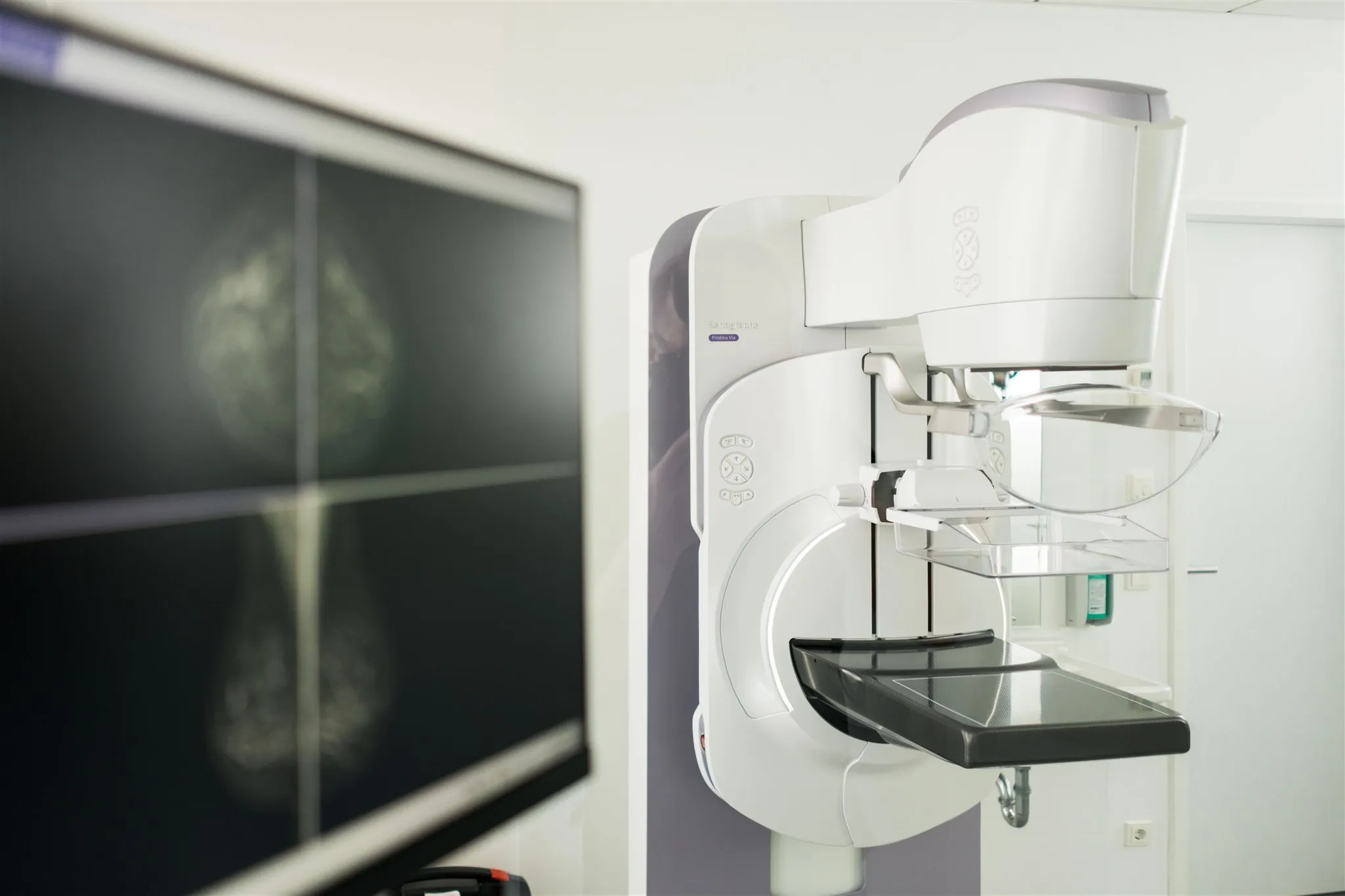
This is the standard procedure of the mammography screening program but also of diagnostic mammography (for complaints). Two images of each breast are usually taken from different angles, which are then presented to the radiologist for evaluation.
Contrast mammography/CESM/KM mammography
This examination is a 2D mammogram in which an iodine-containing contrast medium is administered before the examination. Compared to 2D mammography and 3D mammography, it has a slightly increased radiation risk, but on the other hand, it is also significantly more sensitive to changes in the breast that are associated with increased blood flow. With a few exceptions (so-called low-grade DCIS - a precursor tumor), breast tumours generally have increased blood flow. The sensitivity of KM mammography is therefore comparable to that of MRI, but also offers the advantage of discovering the smallest calcifications, some of which occur in precursors of the tumour. The “BRAID trial” from Great Britain shows comparable sensitivity for the discovery of breast cancer for contrast mammography (CESM) and for magnetic resonance imaging of the breast (breast MRI). KM mammography is used to detect potentially malignant breast changes, to assess the size of tumors, to exclude or detect second tumors either in the same breast or on the other side. In addition, the effect of chemotherapy can also be assessed over time. KM mammography is not possible if silicone implants are present (cosmetic or after tumor removal) or if there is an allergy to iodine-containing contrast medium.
If findings can only be detected using KM mammography or MRI, we can also perform km mammograph-based sampling.
MRI of the mamma/ magnetic resonance imaging of the breast:
We perform this examination using a state-of-the-art 1.5 Tesla MRI scanner. MRI mammography, just like contrast-enhanced mammography, is based on the principle of increased blood flow in altered breast tissue. Breast tumors can generally be clearly identified here. Breast MRI, along with contrast-enhanced mammography, is the most sensitive method for detecting invasive breast cancer. Breast MRI is also performed using a contrast agent containing gadolinium. The examination takes approximately 25 minutes, during which the breast is examined using various measurements. A component of this examination is a dynamic measurement in which contrast agent uptake in various tissues is measured over a period of 5 minutes. The speed of contrast agent uptake in a lesion and its washout from the finding provide clues about the nature of the change (malignant or benign). Breast MRI can detect tumor size, tumor location, and potential secondary tumors. Furthermore, it can be used for follow-up care in patients with implants, particularly when glandular breast tissue has been removed – in such cases, it is generally preferred over other methods. Additionally, silicone implants can be examined to detect or rule out any existing implant damage. In this case, the examination is performed without contrast agent. Generally, there are very strict rules under which breast MRI is covered by statutory health insurance. These include: ruling out recurrence after breast cancer if the finding cannot be clearly clarified by mammography and sonography and the tumor treatment was more than 1 year ago; or searching for a primary tumor in cases of breast cancer metastases that cannot be found by mammography or ultrasound. All other breast MRI examinations are generally performed as self-pay services (IGEL) for individuals with statutory health insurance, especially follow-up examinations after cosmetic procedures (silicone implants for cosmetic reasons). Private health insurance companies usually reimburse the costs of a breast MRI.
We are happy to advise you on the various examination options for your specific case.
Ultrasound/breast ultrasonography
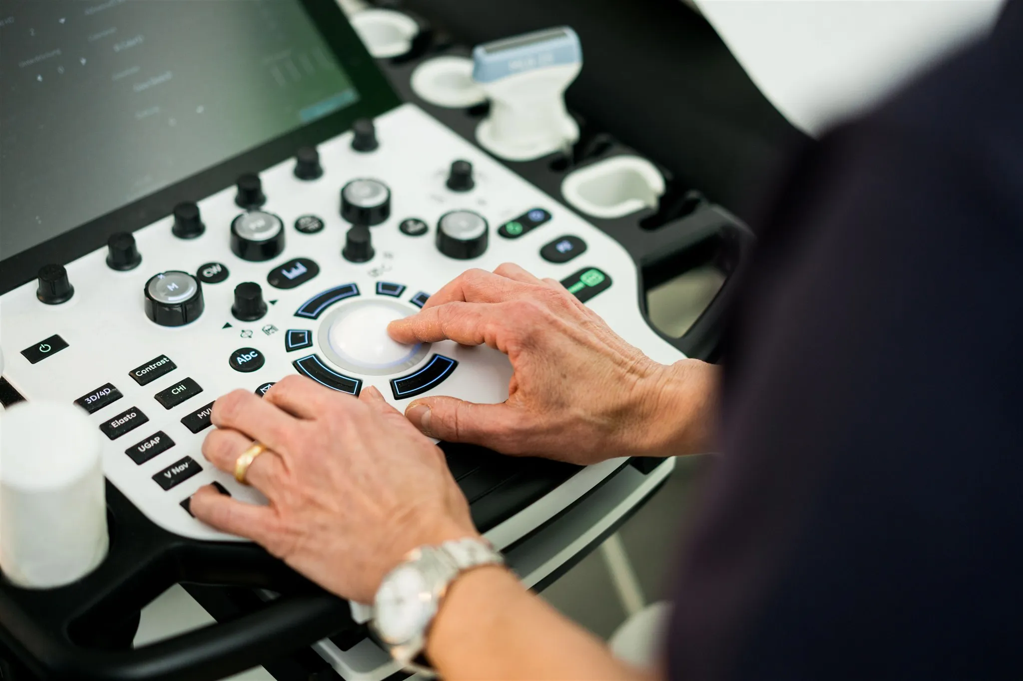
Breast ultrasonography is a standard procedure for clarifying abnormal tactile findings and abnormal findings in mammography. In most cases, it is a supplement to mammography. Using ultrasound waves, the various tissues in the breast are displayed and the doctor interprets what has been seen. If necessary, a sample can then be taken directly from an unusual area under ultrasound control. Ultrasound examination as well as sampling is a service that is covered by statutory and private insurance companies if there is a corresponding indication.
Ultrasound-guided breast biopsy, vacuum suction breast biopsy
In case of abnormal findings, we offer sampling from the affected tissue. This is done in various ways, either under sight using an ultrasound device or under sight using mammography.
In an ultrasound-guided punch biopsy, a needle is guided under local anesthesia under ultrasound vision to the abnormal findings, and small tissue cylinders are then removed. These are then sent to a pathologist for further processing, who evaluates them.
In a vacuum suction biopsy, a needle is presented under X-ray vision, also under local anesthesia, until the findings are found, and small tissue cylinders are then sucked out of the findings with a vacuum. The samples are then also sent to a specialist pathologist for evaluation.
Which type of biopsy we use depends on the findings. If the findings are visible on ultrasound, we remove the tissue under ultrasound vision, if they are only visible on mammography, we perform a vacuum suction biopsy.
As a rule, every finding from which we take a sample is marked with a clip, which, should the findings be surgically removed later, is also removed again. If the findings turn out to be benign and no surgical intervention is required, the clip remains in the chest so that it is clear that a sample has already been taken from this area.
Artificial intelligence
For years and back then, we were one of the first institutes in Germany to support artificial intelligence algorithms to evaluate our images. Various systems are used here, all of which are CE and FDA certified. We use AI algorithms to help identify 2D mammograms, 3D mammography/tomosynthesis, determine mammographic breast density, assess the production quality of automated breast ultrasound, and estimate the risk of being diagnosed with breast cancer within the next two years after the current mammogram. Based on these measurements, we also recommend further measures or individualized examination intervals as part of curative/diagnostic mammography. In mammography screening, the algorithms help us to evaluate the images and find the smallest changes
Computed Tomography
Computed tomography is a procedure for imaging body regions using a rotating X-ray tube that produces cross-sectional images. These are used to detect diseases such as tumors, inflammations, fractures, and others. We use this procedure in our practice for therapy preparation in women diagnosed with breast cancer and for monitoring the progress of these therapies. The scans are predominantly performed with the administration of an iodine-based contrast agent to ensure better assessment. We strive to schedule appointments as quickly as possible so that therapies can be initiated promptly.
preparation on your appointment
To ensure your appointment with us is efficient and pleasant, we've prepared a document to help you have everything ready.
You can easily download all important documents here and fill them out at home before your appointment. Feel free to email us the completed documents beforehand, or bring them printed to your appointment.
Range of services
Additional Services
Regular check-ups are an important step for your health. With modern diagnostics and great care, we help you identify changes at an early stage.

Tomosynthesis/3D mammography
This is a mammogram that takes layered images of your breast. The examination is comparable to that of a 2D mammogram, but is more informative than a 2D mammogram, especially for women with dense mammary gland tissue. This examination is usually covered by private health insurance companies; statutory health insurance companies do not cover these costs, which is why we offer it as an IGEL service. The TOSYMA study carried out as part of the German mammography screening program, in which we were also involved together with the University of Münster, clearly demonstrates the advantage of tomosynthesis over 2D mammography. International studies such as the ReToMo Trial (Italy), the ToBe Trial (Norway) and the MAITA Trial (Italy), in which Dr. Axel Gräwingholt was involved, also impressively demonstrate the advantage of tomosynthesis in detecting tumors
ABUS (automated breast ultrasound)
This is a relatively new method that uses ultrasound waves to generate images of your breast similar to a conventional ultrasound image but in 3 image levels. The difference is that layered images are created which are easier for radiologists to assess and which appear to be superior to manual ultrasound even for very small lesions. Each breast is examined overlapping with an ultrasound probe by a specialist (MTRA or MFA) so that even small changes do not escape. The evaluation is then carried out by the radiologists. This benefit is not a benefit provided by the statutory health insurance funds; it is reimbursed by private insurance companies.
In particular, women with dense mammary gland tissue, who cannot receive additional examinations as part of the mammography screening program due to inconspicuous mammography, or women who have not yet been invited to mammography screening due to their age, benefit from this examination method. Implant ruptures can also be easily presented in an overview using this method. We offer this service as an IGEL service. We would be happy to advise you on the advantages and disadvantages of this process in your specific case and discuss how to proceed.
Partner